The majority of dental pain is an acute response to inflammation. The acute pain associated with dental trauma, infection, or surgery is usually predictably managed with medications. The key in managing pain with medication is to provide a sufficient dose of a particular drug to minimize pain onset and give the patient comfort.
We, at Quality Dental Care also rule out the cause of the dental pain and diagnose with clinical examination and necessary X-Rays (Intra-Oral or OPG) and discuss the required treatment plan.

X-RAYS (Intra-oral and Full mouth)
We have 2 main types of dental x-rays:
- Intraoral (the x-ray film is inside the mouth) and
- Extraoral (the x-ray film is outside the mouth).
Intraoral x-rays are the most common type of x-ray. There are several types of intraoral x-rays. Each shows different aspects of teeth.
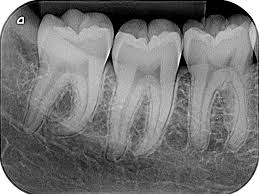
- Bite-wing x-rays show details of the upper and lower teeth in one area of the mouth. Each bite-wing shows a tooth from its crown (the exposed surface) to the level of the supporting bone. Bite-wing x-rays detect decay between teeth and changes in the thickness of bone caused by gum disease. Bite wing x-rays can also help determine the proper fit of a crown (a cap that completely encircles a tooth) or other restorations (eg, bridges). It can also see any wear or breakdown of dental fillings.

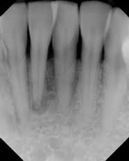
- Periapical x-rays show the whole tooth — from the crown, to beyond the root where the tooth attaches into the jaw. Each periapical x-ray shows all teeth in one portion of either the upper or lower jaw. Periapical x-rays detect any unusual changes in the root and surrounding bone structures.
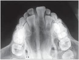
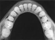
- Occlusal x-rays track the development and placement of an entire arch of teeth in either the upper or lower jaw.
- Extraoral x-rays are used to detect dental problems in the jaw and skull. There are several types of extraoral x-rays.
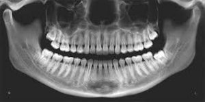
- Panoramic x-rays (OPG – Orthopantomogram) show the entire mouth area — all the teeth in both the upper and lower jaws — on a single x-ray. This x-ray detects the position of fully emerged as well as emerging teeth, can see impacted teeth, and help diagnosis tumors.
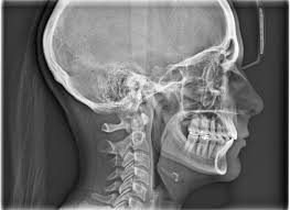
- Cephalometric projections show an entire side of the head. This x-ray looks at the teeth in relation to the jaw and profile of the individual. Orthodontists use this x-ray to develop each patient’s specific teeth realignment approach.